  |
| Welcome > Menu > Module 1 – Understanding Pressure Ulcers > Topic 2: Skin Anatomy and Development of a Pressure Ulcer > Anatomical Changes to Tissue | |||
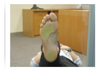
Anatomical Changes to Tissue The picture directly to the right of the screen illustrates the anatomical changes to the tissue as a result of the heel being compressed between the hard surfaces of the glass plate and the bony structures in the heel. Click on the clipboard icon for some key points to consider in relation to the potential damage being caused in these pictures. Click on the Next button to continue.
|
Last updated:
27 March, 2008
This web site is managed and authorised by the Statewide Quality Branch, Rural & Regional Health & Aged Care Services Division of the Victorian State Government, Department of Health, Australia |
|
Copyright | Disclaimer | Privacy statement | State Government of Victoria home | Download help For general enquiries to the Department of Health telephone 61 3 90960000 |