| |
Stage 2 Examples
Examples of different Stage 2 PUs.
Click on each example.
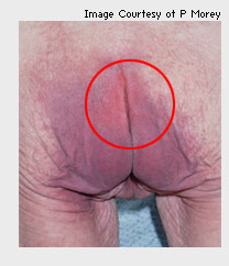
Example A
This illustrates areas of non-blanchable erythema over a wide area but the abrasion near the top of the natal cleft is evidence of epidermal stripping. Hence it is a Stage 2 PU.
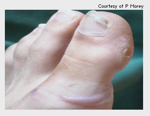
Example BThis slide shows a Stage 2 PU caused by anti-embolic stockings. There is a blister on the lower lateral aspect of the great toe.
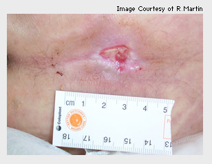
Example CThis slide shows a Stage 2 ulcer presenting as a shallow crater. This presentation is common over areas with underlying bony prominences such as the natal cleft, heel and trochanter. The whiteness around the wound is evidence of moisture being present and is a contributing factor.
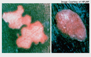
Example DIn darker skin, changes are quite marked due to epidermal stripping (as evidenced by a loss of pigment). The wound on the left is a new injury as it is a pink and shiny wound.
The wound on the right is an older healing injury. Note changes in colour at the margin as epidermis begins to epithelialise across the wound.
Example E
This slide shows separation between the epidermis and dermis as a result of shearing and friction, and leakage of fluid into interstitial spaces. The defining characteristic of this ulcer is the persistence of redness.
All blisters are categorised as Stage 2 irrespective of whether they show signs of necrosis at the perimeters of the blister, or within the blister sac itself.
Click on the Next button to continue.
|