| |
Patient Case Study
Here is a second case study for you to read through to illustrate the impact on an individual patient and a hospital.
George, had a low anterior resection for cancer of the bowel. He developed
an anastomotic leak, peritonitis, abdominal wound dehiscence and multiple
faecal fistulae. He was transferred from a rural hospital to an ICU in
a major metropolitan teaching hospital where he remained for approximately
three months. Click on each of the periods to see how George’s wound developed.
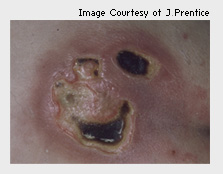
This photo was taken approximately 14 days after admission to ICU. The patient is not being moved as nurses stated it made the patient ‘haemodynamically unstable’. This is a misnomer that must be overcome. This picture shows redness of the surrounding skin, which shows the effects of pressure. The wound itself is necrotic.
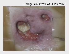
This is a photo taken several days later. You’ll see the use of dressings to debride necrotic tissue and the use of stomahesive to protect peri-wound skin. This picture shows:
- necrotic and sloughy tissue which masks the depth of underlying tissue damage
- less necrotic tissue, indicating improving wound condition. However, the assessor must query what type of tissue is exposed - is the yellow tissue slough, tendon or bone?
- obvious sinus tracking from one wound to the other – there is a need to probe the wound to determine the length and depth of the sinus.
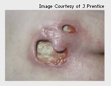
This photo was taken approximately 10 days later. This picture shows:
- merging of the smaller wounds
- depth of tissue loss
- a Stage 4 PU - bone is exposed
- communication between the ulcers is more clearly defined
- the degree of induration in the tissues.
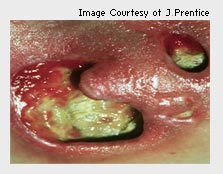
This is a close-up of previous picture. This picture shows:
- induration
- cellulitic skin
- dense, adherent slough which masks what is going on
- probing of the wound identifies underlying bone.
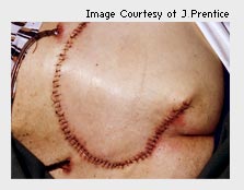
This photo is taken after an extended hospital stay of six weeks after discharge from ICU. The patient has undergone surgery for rotational flap to correct the tissue deficit from the PU. At this point, take a moment to consider the effect on this patient and his family, the loss of income and the costs to the hospital. You may be interested to know that the patient weighed 120kg on admission and was 56kg on discharge.
Click on the Next button to continue.
|